 4.4 Out of 259




 (214) 308-1106
  Book Online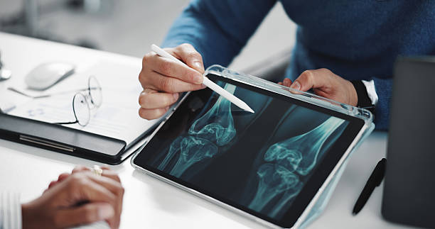
TL;DR
A walk-in clinic with on-site radiography can image bones and chests: suspected fractures of the hand, wrist, elbow, foot, ankle, and ribs, plus chest films for pneumonia and abdominal films for obstruction or stones. Results are usually reviewed during the visit, so people leave with a splint and a plan rather than a referral.
Plain X-ray is a bone-and-air test. It does not show ligaments, tendons, cartilage, or bleeding inside the skull, and a first film can miss a small fracture that only becomes visible a week or two later. Head injuries, open fractures, obvious deformity, numbness, severe chest or abdominal pain, and any injury from a high-speed crash belong in an emergency room, where CT is available.
What a Plain X-Ray Can Actually See
Radiography works on density. The National Institute of Biomedical Imaging and Bioengineering explains that bone contains calcium, which has a higher atomic number than most tissue, so bones absorb the beam and appear whiter against the black background, while fat, muscle, and air-filled lungs travel through more easily and land in shades of gray. That is the National Institute of Biomedical Imaging and Bioengineering’s own X-ray fact sheet, a federal explainer written for patients.
That single physical fact is the whole map of what a walk-in X-ray can and cannot answer.
It answers bone questions well. Is the radius broken? Did the ankle fracture shift out of line? Has that finger dislocated?
A foreign object in the foot, a rib fracture behind the pain, a pattern in the lung fields that fits pneumonia: all of those are inside its range. The FDA’s own description of radiography lists orthopedic evaluation and the detection of foreign objects and structural damage among its ordinary uses.
Soft tissue is where it goes quiet. A completely torn ankle ligament and a badly sprained one look identical on film, because neither shows up. The X-ray in that visit is not there to find the sprain. It is there to prove the bone underneath is intact, which is a real answer and the one that changes whether you get a splint or a cast.
Cartilage, tendons, discs, menisci, and bleeding inside the skull sit in the same blind spot. NIBIB describes CT as combining X-ray with computer processing to build cross-sectional images that are more detailed than plain radiographs, which is precisely why a hospital reaches for it when the question is about what is happening inside soft tissue rather than to a bone.
So the honest framing of a same-day film is narrow and useful. It rules a fracture in or out, it looks at the lungs, and it does that in minutes. Anything requiring a look at the structures between the bones is a different machine in a different building.
Which Injuries Belong in a Walk-In Clinic
Some sorting is easy once you know what the machine is for.
| The injury | Where it usually belongs |
| Rolled ankle, can limp on it, no deformity | Walk-in clinic with X-ray |
| Fall on an outstretched hand, wrist swollen and sore | Walk-in clinic with X-ray |
| Jammed or crooked-looking finger or toe | Walk-in clinic with X-ray |
| Dropped something on a foot, bruised and painful | Walk-in clinic with X-ray |
| Cough and fever, provider wants to check for pneumonia | Walk-in clinic with X-ray |
| Bone visible, skin broken over the fracture | Emergency room now |
| Limb bent at an angle it does not bend at | Emergency room now |
| Head injury with vomiting, confusion, or a blackout | Emergency room now |
| Numb, cold, or gray fingers or toes past the injury | Emergency room now |
| Injury from a highway-speed crash or a big fall | Emergency room now |
When Does an Injury Need an Emergency Room Instead?
An injury needs an emergency room when the answer requires a CT scanner, an operating room, or a specialist within the hour. Walk-in clinics carry plain radiography, not CT and not MRI, so a head injury, a suspected internal bleed, an open fracture, or a limb that has lost circulation is out of scope no matter how good the X-ray suite is.
Head injuries are the clearest case, and the reason surprises people. A skull X-ray is not a lesser version of a head CT.
The American College of Radiology rates skull radiography as “Usually Not Appropriate” across every acute head trauma scenario in its appropriateness criteria, and names non-contrast CT of the head as the study to do when a clinical decision rule says imaging is warranted ACR Appropriateness Criteria, Head Trauma. A film of the skull can miss the bleeding that actually matters. Nobody should be reassured by one.
The other emergency-room categories follow the same logic. Bone through skin carries an infection risk that needs washing out in an operating room. A deformed limb may need to be reduced under sedation. A crash at speed can produce chest and abdominal injuries that plain film simply cannot exclude.
Chest pain that feels like pressure, spreads to the jaw or arm, or comes with sweating and shortness of breath is a cardiac evaluation, not an imaging errand: call 911.
None of this is an argument for skipping care. It is an argument for going to the right building the first time, because the wrong one costs you hours you are already in pain for.
A Clear X-Ray Does Not Always Mean Nothing Is Broken
This is the part that almost no article on same-day imaging tells you, and it is the reason discharge instructions matter as much as the film.
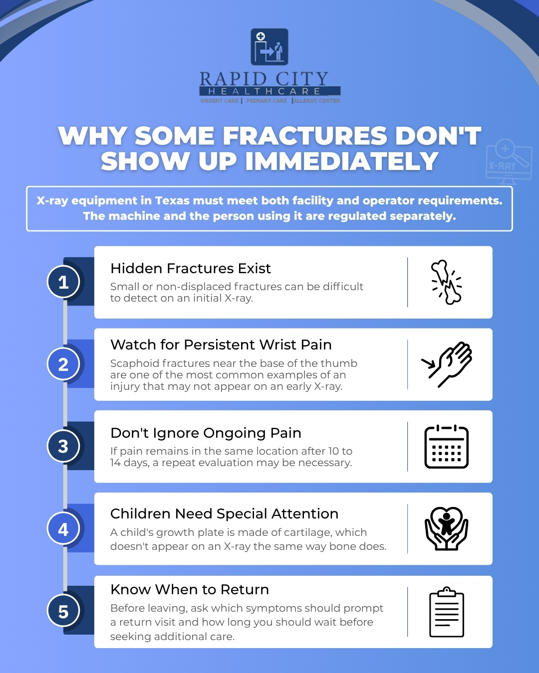
Some fractures are radiographically occult. They exist, they hurt, and they do not show on the first picture because the bone has not shifted and the crack is too fine to change the density the detector sees. The classic one is the scaphoid, a small carpal bone at the base of the thumb that breaks when people catch themselves falling forward. Wrist and hand injuries are where this comes up most.
The same appropriateness criteria series addresses it directly in the American College of Radiology’s document on acute hand and wrist trauma. Radiography is the appropriate first study for that injury, and when those images come back negative or equivocal while clinical suspicion stays high, three next steps are all rated usually appropriate: repeating the radiographs in 10 to 14 days, MRI of the area without contrast, or CT without contrast. The repeat film works because healing bone resorbs at the fracture line and the crack becomes visible when it was not before.
What that means for you, practically, is this. If you leave a walk-in visit with a normal X-ray and a wrist that still hurts in the same specific spot ten days later, the normal X-ray is not the end of the story. Go back. Ask about a repeat film or advanced imaging.
A scaphoid fracture that gets treated as a sprain can go on to a healing problem that is far harder to fix than the original break.
The same principle applies to a child’s growth plate, a stress fracture in a runner’s foot, and a hip in an older adult after a fall. All three are known for quiet first films. A clinician who splints a suspicious wrist despite a clean X-ray is not being cautious for the sake of it. They are treating the exam findings rather than the picture, which is the right order.
Growth plates deserve their own note, because parents hear “the X-ray is clear” and stop worrying. The growth plate is cartilage, and cartilage does not appear on film. A break through it can leave the visible bone looking untouched, which is why swelling and tenderness directly over the growth plate in a child gets splinted and rechecked rather than sent home as a sprain. Children’s ligaments are often stronger than the plate they attach near, so the injury that would sprain an adult ankle can crack a child’s.
Ask two questions before you leave: what specifically should bring me back, and by when. Write the answers down. Ten days later, in the middle of a work week, the memory of a discharge conversation is worth less than a note on the fridge, and the whole value of that repeat-film window depends on somebody acting inside it.
How Much Radiation Is One X-Ray, Really?
A single chest film is one of the smallest doses in diagnostic imaging. The FDA publishes a dose comparison table putting a PA chest X-ray at 0.02 mSv of effective dose, against 2 mSv for a CT of the head, 7 mSv for a CT of the chest, and 8 mSv for a CT of the abdomen FDA, radiation risks from CT. A chest CT is therefore in the region of 350 times the dose of the chest radiograph that might answer the same question.
Extremity films of a hand or foot are smaller still, since the beam is aimed at a limb rather than the trunk.
That does not make imaging free. FDA guidance is that X-ray exams should be performed only when they are needed to answer a medical question or guide treatment, using the lowest reasonable exposure and the minimum number of images, with extra care for children and for anyone who is or might be pregnant. Tell the staff before, not after, if pregnancy is possible.
The stronger argument against an unnecessary film is not the dose anyway. It is that a test taken without a question behind it can turn up something incidental, which leads to another test, then a specialist, then a month of worry, and no benefit. If imaging is offered, it is fair to ask what question it will settle.
Who Is Allowed to Run the Machine in Texas
An X-ray unit in a Texas clinic is a registered and inspected piece of equipment, not an appliance somebody bought.
The facility itself holds a certificate of registration through the Texas Department of State Health Services, whose radiation control program registers medical X-ray machines and sets the technical renewal of that registration at every eight years. Registrants also have to keep written operating and safety procedures under Title 25 of the Texas Administrative Code.
The person positioning you is separately credentialed. The Texas Medical Board certifies medical radiologic technologists and issues limited and temporary permits alongside the general certificate, which is why the staff member running a chest film may hold a narrower scope than a hospital technologist.
Inside the Visit, From Check-In to Answer
The sequence is short, which is the point of walking in.
A provider examines the joint first. Where does it hurt exactly, can you bear weight, is there a pulse past the injury, can you move each finger? That exam decides whether a film is worth taking, and it is also the thing that tells a clinician to splint a wrist despite a clean picture.
Then the imaging. You are positioned so the body part sits between the tube and the detector, usually for two or three views, since a fracture invisible from the front can be obvious from the side. Each exposure lasts a fraction of a second. Digital detectors put the image on a screen in the room within moments rather than sending film to a darkroom.
The provider reviews the images during the visit and explains what they see. In many clinics, the study is also sent for a formal reading by a radiologist afterwards, and you get a call if the second read differs. Treatment starts the same day: a splint, a walking boot, crutches, buddy taping, a sling, pain control, and a referral to orthopedics when the fracture needs one.
That single-stop shape is the practical case for a clinic that keeps an On Site Lab and Same-Day Digital X-Ray, Irving, TX, rather than referring imaging out. It is also why the clinic sits in the gap between a family doctor’s office and a hospital: same-day answers for the middle band of injuries, without the emergency department wait for something that is not an emergency.
One limit is worth stating plainly. A video visit cannot image anything. If the question is whether a bone is broken, it has to be answered in person.
August in Irving Fills the Waiting Room With Ankles
Mid-August in this city is the busiest injury stretch of the year, and the calendar explains why.
Irving ISD’s back-to-school calendar brought students in on Tuesday, August 11, for the 2026-2027 year, with football and marching band already practicing through the first heat-rule change in years. Effective August 1, 2026, the University Interscholastic League’s required heat stress plan has schools taking wet-bulb globe temperature readings within 15 minutes before outdoor activity and every 30 minutes during it. Practices are being shortened, moved earlier, and modified across North Texas because of it.
What lands in a clinic chair from all that is predictable: rolled ankles on grass fields, jammed fingers, wrist injuries from linemen catching themselves, shin pain in kids who went from a summer of nothing to two-a-days, and heat illness that has nothing to do with imaging at all. Cleat-to-cleat contact injuries and the first-two-weeks conditioning surge produce most of it. Getting a walk In sports physical for Irving Students and Athletes done before the season is separate from all of this, but it is the same August rush.
Call the Clinic Before You Drive Over With a Hurt Wrist
Two minutes on the phone saves a wasted trip. Ask whether X-ray is on site or sent out, whether a technologist is there during the hours you are coming, and whether they take your plan, since insurance & self pay options differ by clinic. For a child, ask whether they routinely image kids, because positioning a scared six-year-old is its own skill and pediatric visits run differently.
If the injury is on the emergency list above, skip all of that and go to an ER. If it is not, our Irving team at Rapid City Healthcare can examine it, image it, and splint it in one visit, then hand you clear instructions about what should bring you back. Call ahead and tell us what happened.
This article is general information and cannot account for your history or your exam. If something feels wrong, get it looked at.